
Lump Behind Ear: Causes, Diagnosis and Treatment
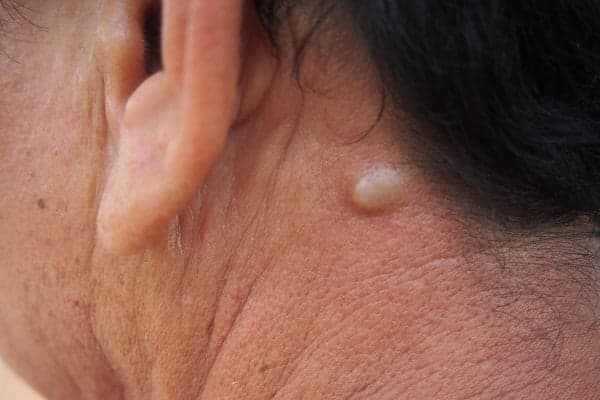
Table of Content
What Causes Lumps Behind the Ear?
Diagnosis of Lumps Behind the Ear
Treatment for Lumps Behind the Ear
When Should You Contact a Doctor?
Conclusion
FAQs
Finding a lump behind the ear is alarming in a way that most minor symptoms are not - the location feels proximate to the brain, and lumps in the head and neck region carry an association with serious pathology that a rash or muscle ache does not. The majority of lumps that develop in this area are entirely benign. Epidermoid cysts, lipomas, reactive lymph nodes, and skin tags account for most presentations. The retro-auricular region is anatomically complex with lymphatic tissue, the mastoid process, the parotid gland, and the posterior auricular nerve all converging here which means that some lumps do represent conditions requiring clinical evaluation and treatment.
Understanding what is most likely and which features distinguish a lump that needs prompt assessment from one that can be monitored is essential.
What Causes Lumps Behind the Ear?
The differential for a retro-auricular lump is broad. These are the most clinically relevant causes:
Lymph node enlargement: The posterior auricular and mastoid lymph nodes drain the scalp, ear, and adjacent skin. They enlarge in response to local infections like otitis media, scalp folliculitis, or infected skin and less commonly in the context of systemic infections such as infectious mononucleosis or haematological malignancy. A soft, tender, mobile node that appears alongside an obvious infection source is almost always reactive and resolves with the underlying illness.
Epidermoid cyst: The most common lump encountered in this region. Epidermoid cysts form when keratin-producing epithelial cells become trapped beneath the skin surface - typically following follicular obstruction or minor trauma. They are smooth, round, and generally non-tender unless infected. A characteristic central punctum may be visible. Most require no treatment unless they enlarge, rupture, or repeatedly infect.
Sebaceous cyst: Frequently conflated with epidermoid cysts clinically, true sebaceous cysts arise from sebaceous gland obstruction rather than epidermal inclusion. The clinical appearance is similar - a soft, mobile subcutaneous swelling and management follows the same principles.
Lipoma: A benign proliferation of mature adipocytes within a fibrous capsule. Lipomas are soft, lobulated, non-tender, and move freely under the skin. They grow slowly and rarely cause symptoms beyond their presence. Excision is straightforward when requested, but the majority do not require treatment.
Skin tags (acrochordons): Small, pedunculated outgrowths of skin arising from areas of friction. The post-auricular sulcus (where the ear meets the scalp) is a common site. Skin tags are entirely benign and require treatment only for cosmetic reasons or if they become irritated.
Mastoiditis: Bacterial infection of the mastoid air cells, typically as a complication of untreated or inadequately treated otitis media. The resulting swelling behind the ear is tender, warm, and often accompanied by anterior displacement of the pinna. This is one presentation that requires urgent assessment as its complications including subperiosteal abscess, meningitis, and sinus thrombosis are serious.
Bone spur (osteophyte): Bony outgrowths on the mastoid process are uncommon but do occur, typically in the context of chronic inflammatory change. They present as hard, fixed lumps adherent to the underlying bone - a clinical feature that distinguishes them from soft tissue causes.
Parotid gland pathology: The parotid gland primarily lies anterior to the ear, but its tail extends posteriorly and can produce a retroauricular swelling when enlarged. Parotid causes include viral parotitis (mumps), calculi causing obstruction, and benign or malignant parotid tumours.
Trauma: Direct injury to the post-auricular region can produce haematoma or localised oedema that presents as a lump. A haematoma following blunt trauma to the mastoid region warrants imaging to exclude underlying fracture.
Diagnosis of Lumps Behind the Ear
Clinical assessment is the starting point. A thorough history including onset, rate of growth, associated pain, preceding infection, systemic symptoms such as fever or weight loss, and relevant medical or family history combined with careful physical examination will identify the probable cause in most cases.
Examination should include the lump itself (consistency, mobility, tenderness, overlying skin changes), the ipsilateral ear canal and tympanic membrane, the regional lymph node chains, and the oropharynx.
Where physical examination is insufficient to establish a diagnosis or where the clinical features raise concern for malignancy, the following investigations are used:
Ultrasound: The first-line imaging modality for soft tissue lumps. Ultrasound distinguishes solid from cystic structures, identifies internal vascularity, and guides aspiration if required. It is widely available, involves no radiation exposure, and provides useful characterisation of most benign causes.
CT or MRI: Cross-sectional imaging is indicated when deep tissue extension is suspected, when the lump has features concerning for malignancy, or when mastoiditis or intracranial involvement needs to be excluded. MRI provides superior soft tissue resolution; CT is faster and better for bony pathology.
Fine needle aspiration cytology (FNAC): A minimally invasive procedure in which a fine needle is used to aspirate cells from the lump for cytological analysis which can characterise lymph node enlargement and cystic lesions & can differentiate reactive from neoplastic lymphadenopathy with reasonable accuracy.
Excision biopsy: Where diagnosis cannot be established through less invasive means, or where complete excision is both diagnostic and therapeutic, surgical removal of the lump with histopathological analysis provides definitive characterisation.
Treatment for Lumps Behind the Ear
Treatment is dictated by the underlying cause. Several options exist across the spectrum from observation to surgery.
Observation: Small, asymptomatic, clearly benign lumps like stable lipomas, incidental epidermoid cysts and skin tags do not require active treatment. A documented baseline with a clear plan for reassessment if the lump changes is the appropriate management for most of these.
Antibiotics: Infected cysts, reactive lymphadenopathy secondary to bacterial infection, and early mastoiditis are managed with oral antibiotics directed at the likely organism. Mastoiditis with subperiosteal abscess formation requires intravenous antibiotics and surgical drainage. Completing the full antibiotic course is essential to negate premature cessation risks (relapse & resistance).
Aspiration: Fluid filled cysts can be drained under ultrasound guidance so that you experience immediate reduction in size and relief of pressure symptoms. Recurrence is common without concurrent removal of the cyst wall (so aspiration is often a temporising measure rather than definitive treatment).
Surgical excision: The definitive treatment for epidermoid cysts, lipomas, and symptomatic skin tags. Complete excision of the cyst wall is necessary to prevent recurrence. For infected cysts, surgery is deferred until the acute infection has resolved. Parotid tumours and lesions suspicious for malignancy require excision by an appropriate surgical specialist.
Radiation therapy: Reserved for confirmed malignant lesions where surgery is not feasible. Sometimes your doctor recommends it as adjuvant treatment following excision of high-risk tumours. This is not a consideration for the majority of retroauricular lumps encountered in clinical practice.
When Should You Contact a Doctor?
Any new lump behind the ear warrants at minimum a clinical assessment, but certain features make prompt review particularly important:
A lump that is growing - particularly one that has doubled in size over weeks rather than months.
Pain, erythema, or warmth at the site, especially if accompanied by fever.
Systemic features: unexplained weight loss, drenching night sweats, or persistent fatigue alongside a lump.
A lump that is hard, fixed to the underlying tissue, and non-tender.
Posterior pinna displacement or otalgia alongside a retro-auricular swelling.
Any new lump in a patient with a personal or family history of cancer, or in a patient who is immunocompromised.
Conclusion
Most lumps that develop behind the ear are benign like epidermoid cysts, lipomas, and reactive lymph nodes and account for the majority of presentations. They are manageable, and in many cases require no active treatment at all.
What matters is accurate identification. The post-auricular region contains structures whose pathology such as mastoiditis, parotid malignancy, and lymphoma carries genuine clinical consequences if missed or delayed. A new lump in this area deserves at least an initial clinical assessment to establish its probable nature, clarify whether investigation is needed, and set appropriate expectations for monitoring or treatment.
FAQs
1. Is swelling of the lymph nodes behind my ear a cause for concern?
Posterior auricular lymph node enlargement is usually reactive - a response to infection in the scalp, ear canal, or adjacent skin - and resolves once the underlying cause is treated. It becomes a concern when there is no identifiable local infection source, when the nodes are firm and non-tender rather than soft and tender, when they continue to enlarge beyond two to three weeks, or when systemic symptoms such as fever and weight loss accompany them.
2. Is it possible that a lump behind the ear is cancerous?
Malignancy is an uncommon cause of retroauricular lumps, but it is not negligible. Lymphoma can present as posterior auricular lymphadenopathy, often alongside involvement of other nodal groups. Parotid malignancy particularly mucoepidermoid carcinoma and adenoid cystic carcinoma can extend to produce a retroauricular mass. Metastatic disease from scalp or cutaneous primary tumours is also possible. Hard, fixed, painless lumps that grow progressively in the absence of infection are the clinical features that raise this concern most strongly, and they should be evaluated without delay.